Image


                        Figure Caption


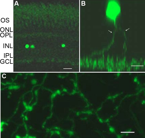
Fig. 4 Morphology of GFP-expressing cells in the retina. A: Localization of GFP-positive cells can be seen in this vertical retinal section. Most of the GFP-expressing cells were found at the proximal cellular row of the inner nuclear layer. Abbreviations: ONL: outer nuclear layer (ONL); OPL: outer plexiform layer (OPL); INL: inner nuclear layer; IPL: inner plexiform layer. B: This z-stack image shows the somata and processes of a single cell. C, GFP fluorescent fiber network in the inner plexiform layer. Scale bar equals 20 μm for A and C, and 10 μm for B.
                
                    
                        Figure Data


                        Acknowledgments


This image is the copyrighted work of the attributed author or publisher, and
ZFIN has permission only to display this image to its users.
Additional permissions should be obtained from the applicable author or publisher of the image.


        