Image


                        Figure Caption


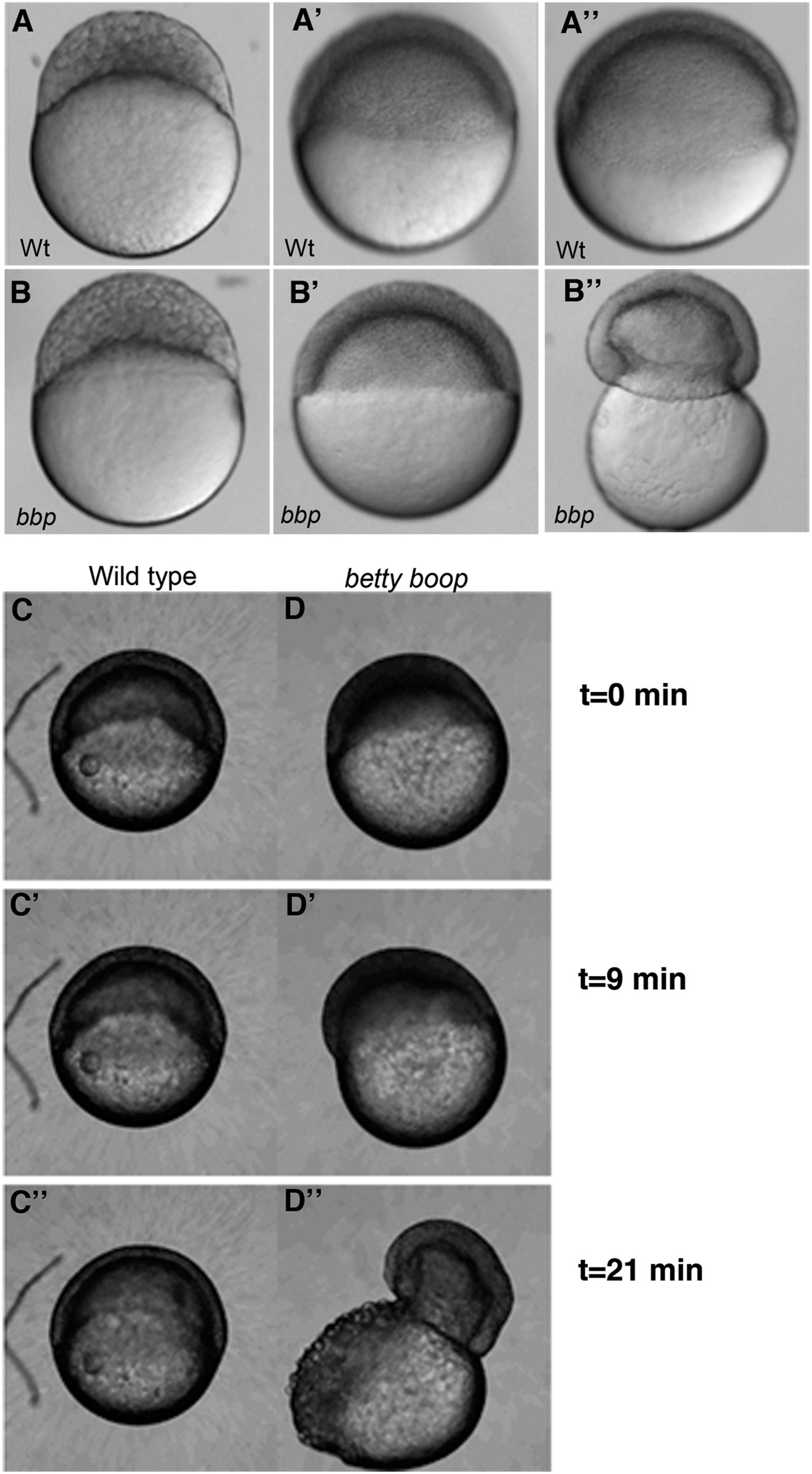
Fig. 2 bbp embryos constrict at the margin at 50% epiboly, bursting the yolk.
Still images of WT and bbp embryos at the 1000-cell stage (A, B), 50% epiboly (A′, B′), and 50% epiboly at the burst (A″, B″). bbp embryos constrict just as they reach 50% epiboly. (C and D) Selected frames from time-lapse movies of WT and bbp embryos showing that the constriction occurs rapidly once the embryos have reached 50% epiboly.
                
                    
                        Figure Data


                        Acknowledgments


This image is the copyrighted work of the attributed author or publisher, and
ZFIN has permission only to display this image to its users.
Additional permissions should be obtained from the applicable author or publisher of the image.
      Full text @ PLoS Genet.