Image


                        Figure Caption


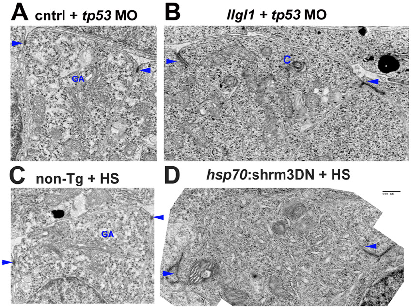
Fig. S3 Apical domains of retinal neuroepithelia. (A-D) Transmission electron microscopy of retinal neuroepithelia from embryos injected with 8 ng control+ 8ng tp53 MO (A), 8 ng tp53 + 8ng llgl1 ATG MO (B), wild-type with heat shock (HS) (C) or carrying the hsp70:shrm3DN:ires:mCherry transgene (D). C and D were also subjected to heat shock (HS) prior to fixation. These images are the non-psuedocolored version from Fig. 3 and Fig. 6. Scale bar: 500 nm. Arrows indicate adherens junctions. GA, golgi apparatus; C, centrosome.
                
                    
                        Figure Data


                        Acknowledgments


This image is the copyrighted work of the attributed author or publisher, and
ZFIN has permission only to display this image to its users.
Additional permissions should be obtained from the applicable author or publisher of the image.
      Full text @ Development


        