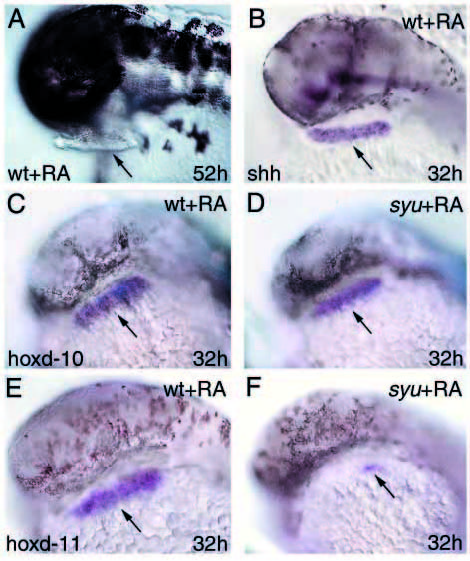
Fig. 5 Effect of retinoic acid treatment on wild-type and syu pectoral fin buds. (A-F) retinoic acid-treated embryos. (A) Embryo at 52 hours; (B-F) whole-mount in situs of embryos at 32 hours of development. (A,B,C,E) Wild-type embryos treated with retinoic acid; (D,F) syu embryos treated with retinoic acid. (B) shh RNA; (C,D) hoxd-10 RNA; (E,F) hoxd-11 RNA. The pectoral fin buds are elongated and located next to the heavily pigmented anterior end of both wild-type and syu embryos treated with retinoic acid (arrows in A to E). Note also that the posterior markers shh and hoxd-11 are expressed throughout the fin bud mesenchyme in wild-type embryos (arrows in B and E), but not in syu embryos treated with retinoic acid, which instead show a patch of hoxd-11 expression at the posterior margin of the fin bud (arrow in F).
                
                    
                        Image


                        Figure Caption


                        Acknowledgments


This image is the copyrighted work of the attributed author or publisher, and
ZFIN has permission only to display this image to its users.
Additional permissions should be obtained from the applicable author or publisher of the image.
      Full text @ Development


        