
Fig. 2
- ID
- ZDB-IMAGE-180529-39
- Genes
- Publication
- Yang et al., 2017 - Expression patterns of zebrafish nocturnin genes and the transcriptional activity of the frog nocturnin promoter in zebrafish rod photoreceptors.
- All Figures
- Figures for Yang et al., 2017
|
Fig. 2
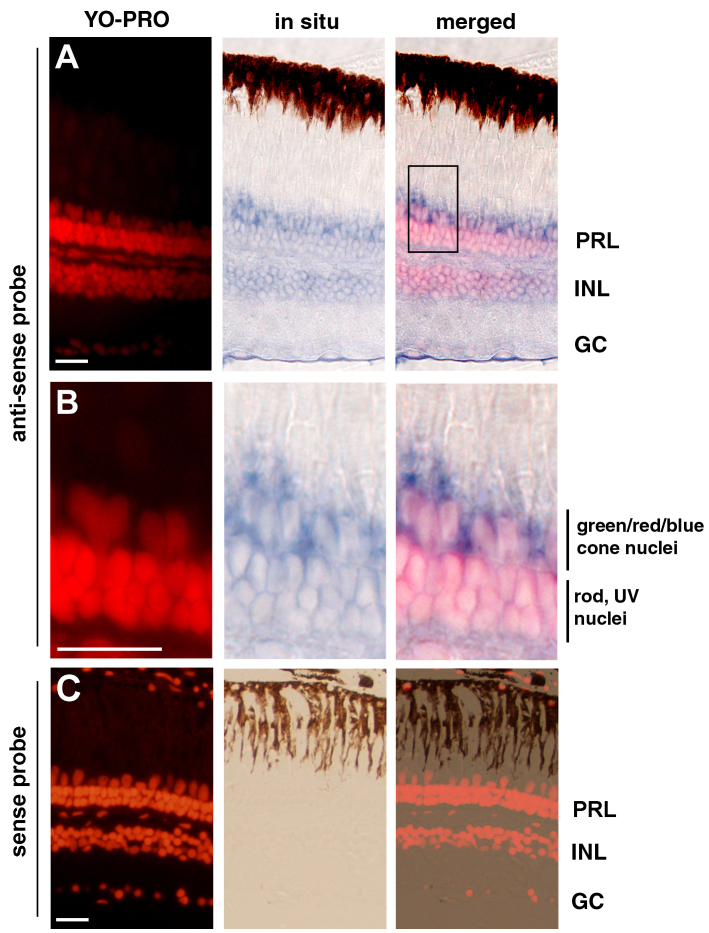
In situ hybridization analysis showed that the nocturnin-b gene is expressed in multiple retinal cell types. A: The nocturnin-b mRNA signals, visualized with an anti-sense probe (blue), were stronger in the photoreceptor layer (PRL) than in the inner nuclear (INL) and the ganglion cell layer (GC). B: A local region of the photoreceptor layer in panel A (boxed region) is magnified to better illustrate the enrichment of the nocturnin-b mRNA signals at the inner segment regions. The cell nuclei were stained with YO-PRO (red). C: In situ hybridization with a sense probe for nocturnin-b did not stain the retina, supporting the staining specificity by the anti-sense probe in panels A and B. Scale bars, 20 μm.