Image


                        Figure Caption


Fig. S1
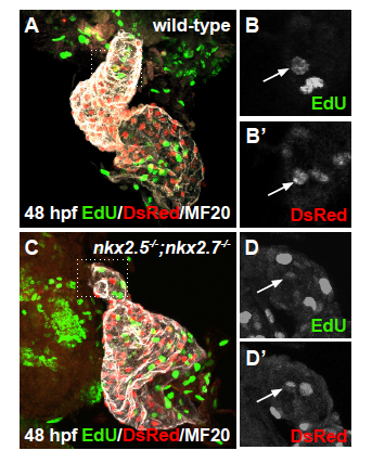
Proliferation of differentiated arterial pole CMs is not affected (A-D) Ventral view, anterior to the top, of the arterial pole region at 48 hpf. Confocal projections of immunohistochemistry for EdU (green), DsRed (red), and MF20 (gray) in wild-type (A,B-B’) and nkx2.5-/-;nkx2.7-/- (C,D-D’) embryos carrying Tg(- 5.1myl7:nDsRed2) following EdU incubation at 24 hpf. Representative images highlight the detection of proliferating nuclei in the OFT myocardium.
                
                    
                        Figure Data


                        Acknowledgments


This image is the copyrighted work of the attributed author or publisher, and
ZFIN has permission only to display this image to its users.
Additional permissions should be obtained from the applicable author or publisher of the image.
      Full text @ Development