
|
Fig. S2
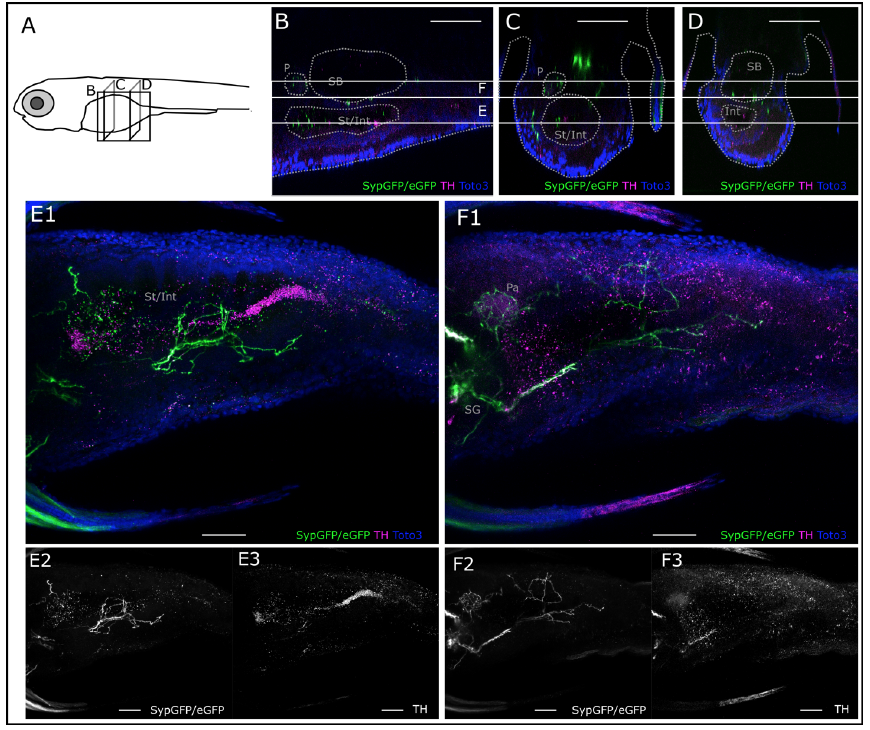
Catecholaminergic innervation of the abdomen, ventral scan. A. Schematic of 6dpf zebrafish larva with scanned sections as depicted in B-D. B. Sagittal section from XZ-orthoslice at 272 μm of z-stack (ventral scan, step size: 1μm, total depth: 390 μm, total width: 509 μm) of larval region including the gut. Dotted grey lines outline anatomical landmark structures, probably the pancreas (P) swim bladder (SB) and stomach and intestine (St/Int). Solid white horizontal lines indicate borders of regions used for MIPs shown in E and F. C. Transverse section from YZ-orthoslice at 100μm of same z-stack. Outlined structures: P and St/Int. D. Transverse section from YZ-orthoslice at 381μm of same z-stack. Outlined structures: SB and Int. E. MIP from subregion as indicated in B-D (slices 170-230). Catecholaminergic projections between and around the SB and the Int/St are labeled with GFP driven by th:Gal4-VP16 and are faintly THimmuno- reactive. F. MIP from subregion as indicated in B-D (slices 230-270). Catecholaminergic projection around the SB is labeled with GFP and anti-TH. Round structure rostral to SB shows strong GFP and anti-TH labeled innervation, probably representing innervation of the pancreas (P) by sympathetic projections. All scale bars: 50μm. GFP: green, panels 2, anti-TH: magenta, panels 3, TOTO-3: blue.