
Fig. 1
- ID
- ZDB-IMAGE-200424-40
- Genes
- Publication
- Guglielmi et al., 2020 - Temporal control of Wnt signaling is required for habenular neuron diversity and brain asymmetry
- All Figures
- Figures for Guglielmi et al., 2020
|
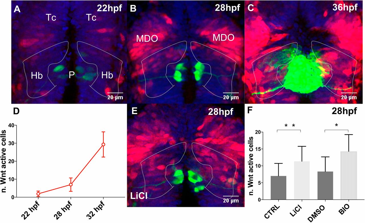
Fig. 1 Wnt signaling activity in and around the developing habenulae. (A-C,E) Projections of confocal z-stacks, dorsal views, anterior is towards the top focused onto the diencephalon of tg(7xtcf-Xla.Siam:nls-mCherry); tg(flh:GFP); tg(foxD3:GFP) transgenic embryos at stages indicated. Nuclei are blue, Wnt active cells are red and the pineal complex is green. Dotted lines highlight the region of the developing habenulae. (D) Graph shows the number of Wnt-active habenular precursors, which are (F) increased, when Wnt signaling is activated by drug treatments, as indicated. Hb, habenulae; MDO, mid-diencephalic organizer; P, pineal; Tc, telencephalon.