IMAGE

Figure 3—figure supplement 1.

ID
ZDB-IMAGE-201012-65
Publication
Inaba et al., 2020 - Keratins and Plakin family cytolinker proteins control the length of epithelial microridge protrusions
All Figures
Figures for Inaba et al., 2020
Image
Figure Caption

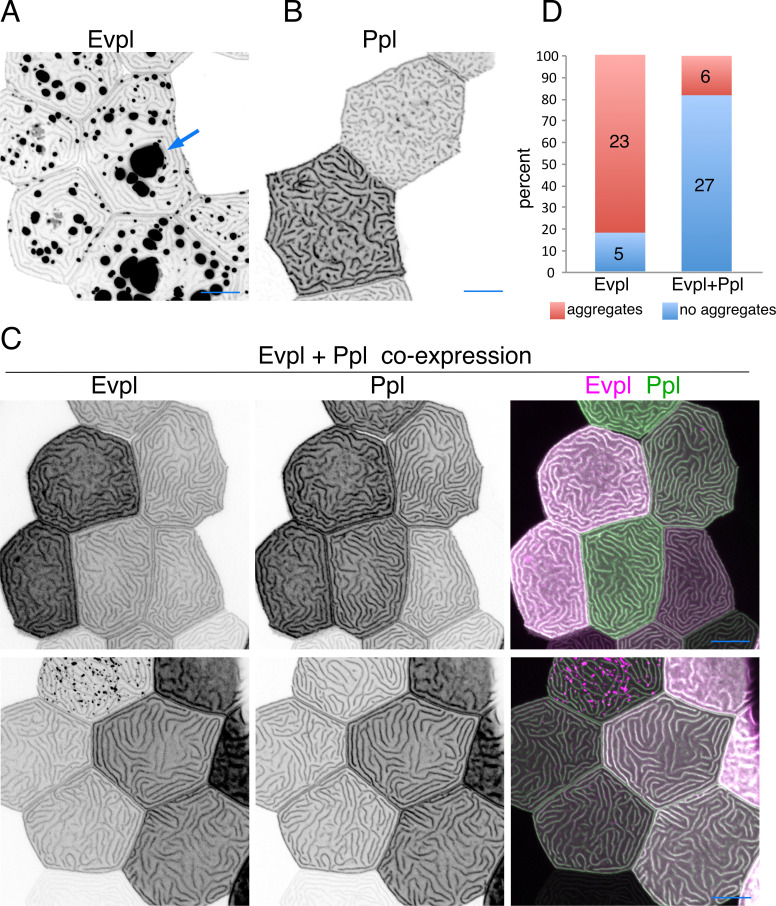
Figure 3—figure supplement 1. Evpl associates with Ppl in microridges.

(A) Evpl-mRuby[BAC] expression in periderm cells of 48hpf zebrafish showing localization to aggregates (arrow) and microridges. Image is a zoomed out version of the image used in Figure 3A. (B) Ppl-GFP[BAC] expression in periderm cells of 48hpf zebrafish showing localization to microridges. (C) Projections of periderm cells in two different fish (each row) co-expressing Evpl-mRuby[BAC] and Ppl-GFP[BAC] at 48hpf. (D) Proportion of cells with aggregates in Evpl- or Evpl/Ppl-expressing cells. Black-and-white images were inverted so that high-intensity fluorescence appears black and low-intensity fluorescence is white. Scale bars: 10 µm.

Acknowledgments
This image is the copyrighted work of the attributed author or publisher, and ZFIN has permission only to display this image to its users. Additional permissions should be obtained from the applicable author or publisher of the image. Full text @ Elife