IMAGE

Figure 4—figure supplement 3.

ID
ZDB-IMAGE-201012-70
Publication
Inaba et al., 2020 - Keratins and Plakin family cytolinker proteins control the length of epithelial microridge protrusions
All Figures
Figures for Inaba et al., 2020
Image
Figure Caption

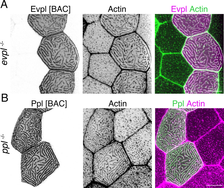
Figure 4—figure supplement 3. Plakin BAC reporters rescue microridge development in plakin mutants.

(A) Lifeact-GFP-expressing cells mosaically expressing Evpl-mRuby[BAC] in periderm cells. Microridges were rescued by BAC expression (compare to neighboring control cells lacking mRuby). These and subsequent black-and-white images were inverted so that high-intensity fluorescence appears black and low-intensity fluorescence is white. (B) Lifeact-mRuby-expressing cells mosaically expressing Ppl-GFP[BAC] in periderm cells. Microridges were rescued by BAC expression (compare to neighboring control cells lacking GFP). Scale bar: 10 µm.

Acknowledgments
This image is the copyrighted work of the attributed author or publisher, and ZFIN has permission only to display this image to its users. Additional permissions should be obtained from the applicable author or publisher of the image. Full text @ Elife