IMAGE

Figure 5.

ID
ZDB-IMAGE-231031-20
Source
Figures for Liu et al., 2023
Image
Figure Caption

Figure 5.

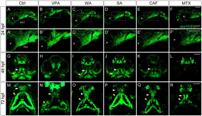
Changes in the morphology of craniofacial anomalies in teratogen-treated sox10:EGFP embryos. (A–F) Lateral live view of the first pharyngal arch (PA1) at 24 hpf. (A′–F′) Enlarged view of the white rectangle in panel A–F. Abnormal morphology of the PA1 was observed in the teratogen-treated embryos. (G–L) The developing palates (white arrowheads) and mandibles (yellow arrowheads) were disrupted in the teratogen-treated embryos at 48 hpf. (M–R) The PA1 defects resulted in a small-sized palate with clefting (cleft palate) and shortening in the Meckel’s cartilage at 72 hpf. e: eye. Scale bar: 100 µm.

Acknowledgments
This image is the copyrighted work of the attributed author or publisher, and ZFIN has permission only to display this image to its users. Additional permissions should be obtained from the applicable author or publisher of the image. Full text @ Toxicol. Sci.