IMAGE

Fig 4

ID
ZDB-IMAGE-241101-23
Source
Figures for Meserve et al., 2024
Image
Figure Caption

Fig 4 
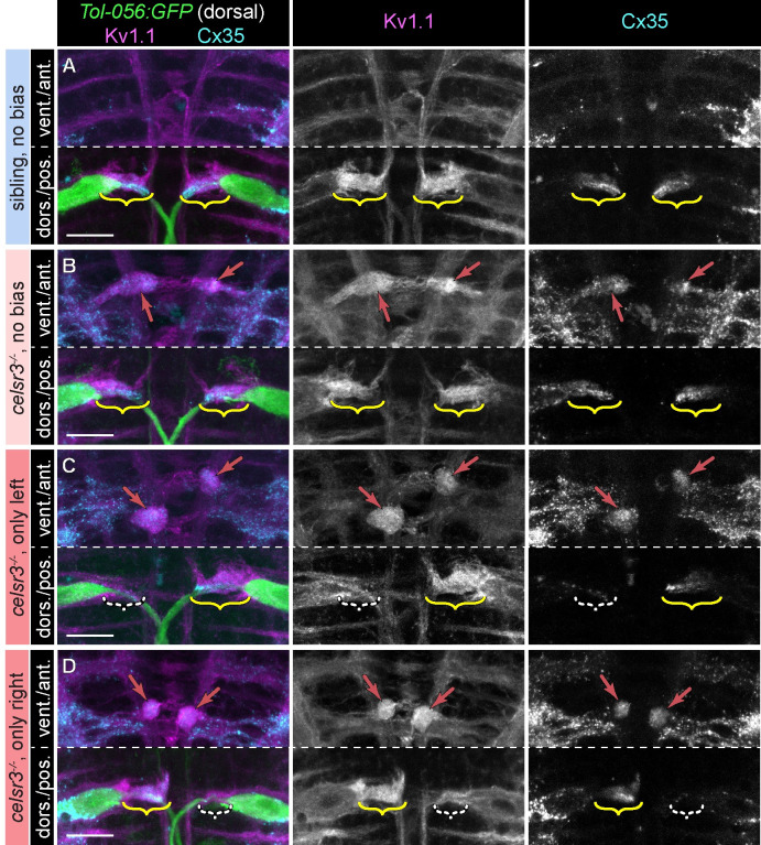
Asymmetric spiral fiber input to Mauthner neurons in celsr3 mutants correlates with biased escapes.

A-D) Region of hindbrain with Mauthner and spiral fiber neurons from 5 dpf Tol-056:GFP larvae in (A) siblings with unbiased escapes (40–60% right/left C1-bend direction ratio; all categories >6 escapes over 20 stimuli), (B) celsr3 mutants with unbiased escapes, (C) celsr3 mutants that only made left C1-bends (right Mauthner predicted active), and (D) celsr3 mutants that only made right C1-bends (left Mauthner predicted active). Composite images are Z-projections with ventral range for anterior portion and dorsal range for posterior portion. Kv1.1 (left and middle panels) localizes to endogenous axon caps (brackets) and ectopic caps (red arrows in B-D) presumed to represent spiral fiber misprojections. α-Cx35 (left and right panels) labels spiral fiber synapses at endogenous (A-D) and ectopic (B-D) caps. White dashed brackets in (C,D) indicate the axon cap with reduced Kv1.1/Cx35 signal, which corresponds to the inactive Mauthner, based on escape bias. Scale bar = 20 μM.

Acknowledgments
This image is the copyrighted work of the attributed author or publisher, and ZFIN has permission only to display this image to its users. Additional permissions should be obtained from the applicable author or publisher of the image. Full text @ PLoS Genet.