IMAGE

FIGURE 2

ID
ZDB-IMAGE-241213-43
Genes
Source
Figures for Chan et al., 2024
Image
Figure Caption

FIGURE 2

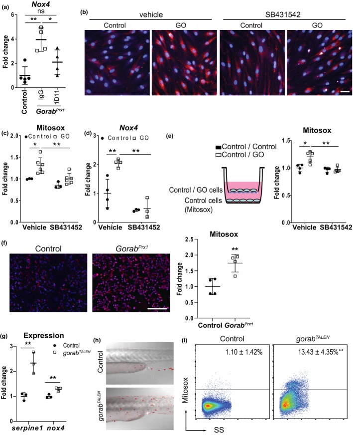
Elevated TGF‐β signaling caused increased Nox4 expression and elevated oxidative stress in GO. (a) 1D11 treatment inhibited the Nox4 upregulation in GorabPrx1 mice (N = 5) (b) Representative images and (c) quantitation of Mitosox intensity in GO patient fibroblasts (N = 3–6) treated with TGF‐β inhibitor SB431542. (d) SB431542 reduced Nox4 expression in GO patient fibroblasts (N = 3–4). (e) Coculturing of control fibroblasts with GO patient fibroblasts caused upregulation of mitosox signal in the control fibroblasts, which can be prevented by SB431542 (N = 4–5). (f) Increased in mitosox signal in the bone marrow of tibiae of 4 weeks old GorabPrx1 (N = 4) in vivo. (g) Knockdown of gorab in zebrafish by TALEN resulted in upregulation of serpine1 and nox4 expression. (h) Representative picture and (i) quantitation of Mitosox positive cells by flow cytometry in gorabTALEN zebrafish embryos. *p < 0.05, **p < 0.01, ns, no statistically significant difference (unpaired Welch's t‐test). Scale bar = 100 μm.

Figure Data
Acknowledgments
This image is the copyrighted work of the attributed author or publisher, and ZFIN has permission only to display this image to its users. Additional permissions should be obtained from the applicable author or publisher of the image. Full text @ Aging Cell