
|
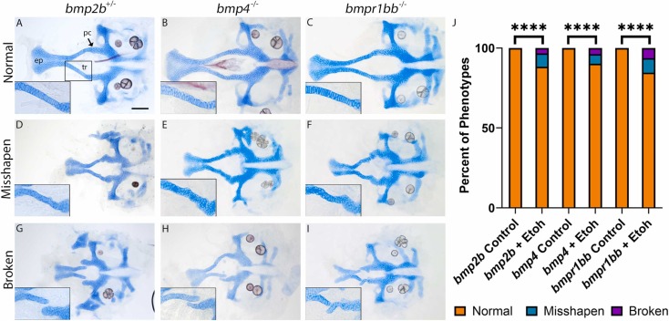
Fig. 1 Multiple members of the Bmp pathway exhibit ethanol-induced palate defects. (A-I) Flat-mount images of neurocranium of 5 dpf larva. Cartilage is blue and bone is red (anterior to the left, scale bar: 100 μm, ep = ethmoid plate, tr = trabeculae and pc = polar cartilage, large images were captured at 10x, inset is 20x image of the left trabecula). (J) total percentage of palate phenotype in ethanol-treated Bmp mutants and controls. (A-C) heterozygous bmp2b or homozygous bmp4 or bmpr1bb mutant larva develop normal neurocranium. Insets show normal stacking of cells in the trabecula. (D-I) Exposure of 1 % ethanol results in a range of defects to palate, from misshapen trabecula (D-F) to breaks in the trabeculae at the polar cartilages (G-I). Percent of phenotypes were quantified and compared for untreated and ethanol-treated larvae for each Bmp component using Fisher’s Exact Test (**** = p <0.0001) (J). “n” for each Bmp component listed in Table 1.
Reprinted from Reproductive toxicology (Elmsford, N.Y.), 131, Lovely, C.B., Bone Morphogenetic Protein signaling pathway - ethanol interactions disrupt palate formation independent of gata3, 108754, Copyright (2024) with permission from Elsevier. Full text @ Reprod. Toxicol.