
|
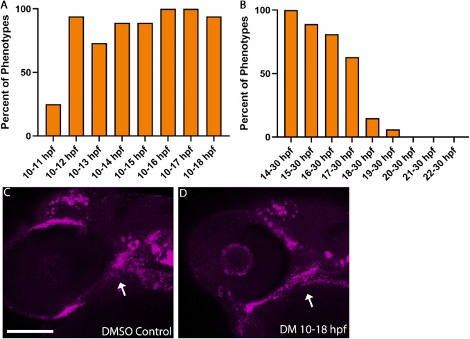
Fig. 5 Dorsomorphin-treated larvae show palate defects but normal gata3 expression when treated between 10 and 18 hpf. (A) Comparison of palate defects in DM-treated larvae with expanding DM exposure window from 10 to 11 hpf out to 10–18 hpf. We observed a large number of palate defects with a DM exposure window as short as 2 hours, 10–12 hpf with increasing duration of exposure resulting in consistent palate defects. (B) Comparison of palate defects in DM-treated larvae with increasingly later initiation time points of DM, 14 hpf (consistent with the latest exposure initiation of ethanol at a higher penetrance (Fig. 4A) out to 30 hpf. Consistent with the ethanol sensitivity exposure window, the percentage of palate defects decreases drastically when DM exposure is initiated at 18 hpf. “n” for each treatment group listed in Table 7. (C,D) Whole-mount, confocal images of DM-treated (10–18 hpf) or DMSO control embryos fluorescently labeling gata3 gene expression at 36 hpf (lateral views, anterior to the left, scale bar: 100 μm). Arrows show normal expression of gata3 in the maxillary domain of the NCC in DMSO and DM-treated embryos as well as ethanol-treated wild type embryos (n = 7 embryos per treatment group).
Reprinted from Reproductive toxicology (Elmsford, N.Y.), 131, Lovely, C.B., Bone Morphogenetic Protein signaling pathway - ethanol interactions disrupt palate formation independent of gata3, 108754, Copyright (2024) with permission from Elsevier. Full text @ Reprod. Toxicol.