
|
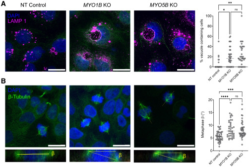
Fig. 3 MYO1B loss impairs membrane recycling and spindle orientation (A) Confocal sections of NT control, MYO1B KO, and MYO5B KO Caco-2 3D cultures stained for the lysosome marker LAMP1 and DAPI, and quantification of the percentage of vacuoles containing cells (Caco-2 NT n = 22, MYO1B KO n = 35, MYO5B KO n = 35). Scale bars, 20 μm. (B) Confocal sections of NT control and MYO1B KO Caco-2 3D cultures stained for β-tubulin and DAPI. The β angle between the spindle axis and the substratum in the confocal x-z dimension was quantified in metaphase (Caco-2 NT n = 42, MYO1B KO n = 40, MYO5B KO n = 44). Scale bars, 20 μm. Data are presented as median; one-way ANOVA test, ∗p < 0.05, ∗∗p < 0.01, ∗∗∗p < 0.001, ∗∗∗∗p < 0.0001.