
|
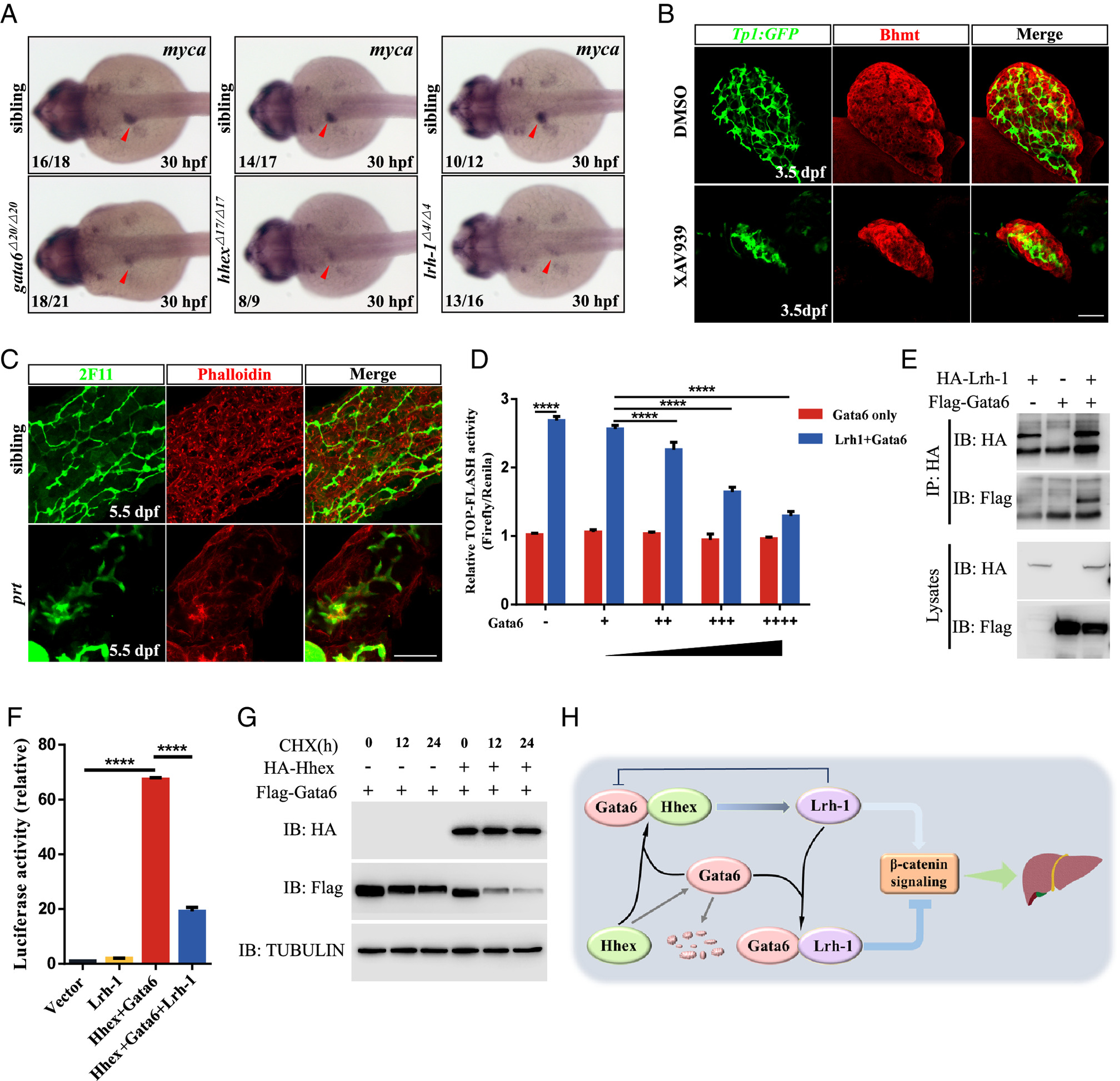
Fig. 5 The coordinated interactions among Gata6, Hhex, and Lrh-1 modulate the output of β-catenin signaling. (A) WISH showing myca expression at 30 hpf. Red arrowhead, liver bud. (B) Immunostaining of Tg(Tp1:GFP) embryos with or without XAV939 treatment (18 hpf-54 hpf) at 3.5 dpf using Bhmt antibody (red, hepatocytes) (n = 10/10, DMSO, n = 11/14 XAV939). (Scale bar, 50 μm.) (C) Immuno- and dye staining of livers in the siblings (n = 14) and prt mutants (n = 12) at 5.5 dpf. (Scale bar, 50 μm.) (D) TOP-FLASH assays wherein Lrh-1 and the increasing amounts of Gata6 expression in HEK293T cells. (E) Coimmunoprecipitation assays of Gata6 and Lrh-1 in HEK293T cells. (F) Luciferase assays of the lrh-1 promoter with Gata6, Hhex, and Lrh-1 in HepG2 cells. (G) Immunoassay of Flag-Gata6 and HA-Hhex with or without CHX treatment at the indicated time points in HEK293T cells. (H) A model to depict the molecular mechanism underlying liver development in zebrafish. Data are mean ± SD, ****P < 0.0001, two-tailed unpaired t test.