
|
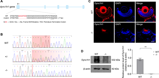
Fig. 1 Identification of dync1h1 knockout zebrafish. (A) Schematic representation of the genomic region and exon/intron structure of dync1h1 in zebrafish. Knockout of dync1h1 results in a truncated protein, with deletion located in exon 1. (B) Direct sequencing comparison of WT, dync1h1+/−, and dync1h1−/− zebrafish lines. (C) Immunofluorescence staining showing dync1h1 protein expression in 5 dpf WT and dync1h1−/− zebrafish. Nuclei are stained with DAPI. (D) Western blot analysis of dync1h1 protein expression in 5 dpf WT and dync1h1−/− zebrafish, with β-actin as the endogenous control. +/−, dync1h1 heterozygote; −/−, dync1h1 homozygote. Scale bar = 50 µm. Data are shown as mean ± SD. **, P < 0.01, n = 3 biologically independent samples per group. DAPI, 4′,6-diamidino-2-phenylin-dole; dpf, days post fertilization; SD, standard deviation; WT, wild type.