
|
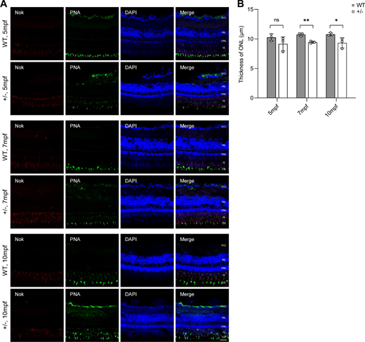
Fig. 4 Mild but progressive retinal degeneration in adult dync1h+/− zebrafish. (A) Immunofluorescence-stained cryosections at indicated ages showing the PRC IS/OS stained with anti-Nok and PNA; nuclei stained by DAPI. (B) Quantitative analysis of ONL thickness in WT and dync1h1+/− retinas over time. +/−, dync1h1 heterozygote. Scale bar = 20 µm. Data are shown as mean ± SD. Not significant (ns), P > 0.05, *, P < 0.05; **, P < 0.01, n = 3 biologically independent samples per group. DAPI, 4′,6-diamidino-2-phenylin-dole; IS/OS, inner segment/outer segment; Nok, nagie oko; ONL, outer nuclear layer; PNA, peanut agglutinin; PRC, photoreceptor cell; SD, standard deviation; WT, wild type.