Image


                        Figure Caption


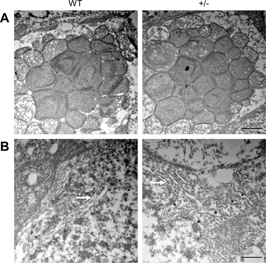
Fig. 6 Prioritization of ER impairment in dync1h1+/− retinas at 20 mpf. (A) TEM images showing well-developed mitochondria in both WT and dync1h1+/− retinas. (B) TEM image highlighting well-developed Golgi (white arrow) in both WT and dync1h1+/− retinas. The black asterisks point to abnormally swollen ER scattering in IS. +/−, dync1h1 heterozygote. Scale bar = 1 µm (A), 500 nm (B). ER, endoplasmic reticulum; mpf, months post fertilization; WT, wild type.
                
                    
                        Acknowledgments


This image is the copyrighted work of the attributed author or publisher, and
ZFIN has permission only to display this image to its users.
Additional permissions should be obtained from the applicable author or publisher of the image.
      Full text @ Invest. Ophthalmol. Vis. Sci.


        