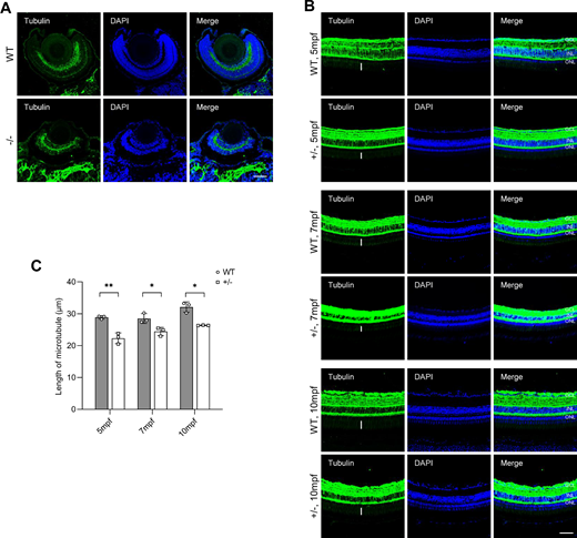
Fig. 7 Dync1h1-deficiency impairs cilium biogenesis. (A) PRC cilium biogenesis in WT and dync1h1−/− retinas at 5 dpf, detected by immunofluorescence for axonemal microtubules using anti-α-acetylated tubulin antibody; nuclei stained by DAPI. (B) PRC development in WT and dync1h1+/− retinas at indicated ages, with axonemal microtubules by anti-α-acetylated tubulin; nuclei stained by DAPI. The lengths of microtube were labeled with white vertical lines. (C) Quantitative analysis of microtubule length in WT and dync1h1+/− retinas across ages. +/−, dync1h1 heterozygote; −/−, dync1h1 homozygote. Scale bar = 50 µm. Data are shown as mean ± SD. *, P < 0.05; **, P < 0.01, n = 3 biologically independent samples per group. DAPI, 4′,6-diamidino-2-phenylin-dole; dpf, days post fertilization; PRC, photoreceptor cell; SD, standard deviation; WT, wild type.
                
                    
                        Image


                        Figure Caption


                        Acknowledgments


This image is the copyrighted work of the attributed author or publisher, and
ZFIN has permission only to display this image to its users.
Additional permissions should be obtained from the applicable author or publisher of the image.
      Full text @ Invest. Ophthalmol. Vis. Sci.


        