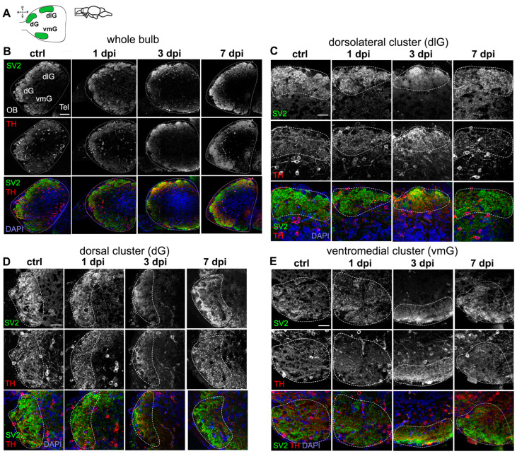
Dysregulation of synaptic contacts in the OB caused by 6-OHDA. (A) Schematic diagram of a sagittal view of the OB, indicating three glomerular clusters: dorsal (dG), dorsolateral (dlG), and ventromedial (vmG). (B–E) Double immunohistochemistry of synaptic vesicle protein 2 (SV2) and tyrosine hydroxylase (TH) in sagittal sections of the OB from controls, 1 dpi, 3 dpi, and 7 dpi groups. Representative images taken from 5–7 animals analyzed per group. Dotted lines indicate the glomerular clusters selected. Whole bulb (B), and magnified views of: (C) dlG cluster, (D) dG cluster, and (E) vmG cluster. Green: Tbr2a; red: TH; blue: DAPI. Scale bars: 100 µm in (B); 50 µm in (C–E).
Acknowledgments
This image is the copyrighted work of the attributed author or publisher, and
ZFIN has permission only to display this image to its users.
Additional permissions should be obtained from the applicable author or publisher of the image.
Full text @ Int. J. Mol. Sci.
Your Input Welcome
Thank you for submitting comments. Your input has been emailed to ZFIN curators who may contact you if
additional information is required.
Oops. Something went wrong. Please try again later.