IMAGE

Fig. 4

ID
ZDB-IMAGE-250703-21
Source
Figures for Trindade et al., 2025
Image
Figure Caption

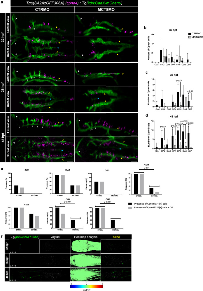
Fig. 4 MT3 regulates Cpne4+ cells during hindbrain vasculature development.

a Maximum projection of the hindbrain vasculature structures in double transgenic zebrafish embryos (Tg(kdrl:CaaX-mCherry);Tg(gSA2AzGFF306A)) immunostained against mCherry (green) and GFP (Cpne4+ cells, magenta) are shown. In CTRMO zebrafish embryos, Cpne4/GFP(+) cells were in close contact with the central arteries (CtAs), while in MCT8MO zebrafish embryos some Cpne4/GFP(+) cells were lost. The white arrowhead indicates the mid-cerebral vein (MCeV), the yellow arrowhead indicates the primordial hindbrain channels (PHBC), and the blue arrowhead indicates the basilar artery (BA). White numbers 1 – 7 indicate the developed CtA in its respective rhombomere. The number of Cpne4/GFP(+) cells between CTRMO and MCT8MO zebrafish embryos was analyzed at b) 32 hpf (n = 9, 8 (CTRMO, MCT8MO)), c) 36 hpf (n = 5, 6 (CTRMO, MCT8MO)) and d) 48 hpf (n = 7, 10 (CTRMO, MCT8MO)); Unpaired t-test; Error bars represent standard deviation. e Graphical view of the correlation between the presence of Cpne4/GFP(+) cells and the presence of each CtA in CTRMO and MCT8MO at 48 hpf shows that a correlation between these two conditions exists for CtAs 4, 6 and 7. Fisher’s exact test. n = 7 (CTRMO), 11 (MCT8MO). For detailed statistics, see Supplementary Data 2. f Dorsal view of the hindbrain of Tg(gSA2AzGFF306A) zebrafish embryos at 32, 36 and 48 hpf after WISH for vegfaa (white) and immunostained against GFP (Cpne4 cells, green) are shown. Heatmap colocalization analysis was used using normalized mean deviation product (nMDP) values and a combined image between GPF and vegfaa expression is shown. Color bar chart indicating no colocalization (-1, blue color) to colocalization (1, red color). Scale bar: 50 μm.

Acknowledgments
This image is the copyrighted work of the attributed author or publisher, and ZFIN has permission only to display this image to its users. Additional permissions should be obtained from the applicable author or publisher of the image. Full text @ Commun Biol