IMAGE

Fig. 10

ID
ZDB-IMAGE-250703-27
Source
Figures for Trindade et al., 2025
Image
Figure Caption

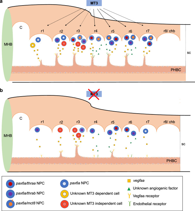
Fig. 10 Proposed model for MT3 Mct8-mediated hindbrain development in zebrafish embryogenesis.

a Illustration of the zebrafish hindbrain with proposed action of MT3 signaling through Mct8 on CtA development. The different pax6a neural progenitor cells (NPC) identified in the different rhombomeres with the TH machinery mct8, thraa and thrab are illustrated. In rhombomere 1 we suggest the involvement of another central nervous system (CNS) cell type that is MT3 dependent and the source of vegfaa required for CtA development. In rhombomere 3, pax6a NPCs are present, but MT3 signaling is not involved in CtA 3 development (dashed arrow line) but enhances its development. Vascularization of rhombomeres 4, 5 and 6 suggests that, besides vegfaa, another angiogenic factor is involved in the angiogenic sprouting of these CtAs. b Illustration of a lateral view of the zebrafish hindbrain showing the effect of Mct8 knockdown and MT3-impaired signaling. The most frequently developed CtAs in the hindbrain of 48 hpf zebrafish embryos are represented. Although significantly reduced in rhombomere 2, vegfaa signaling is still present in MCT8MO zebrafish embryos, showing that another CNS cell type, independent on MT3 signaling, is responsible for releasing vegfaa or able to compensate for the lack of pax6a + /MT3-responsive cells. The inability to rescue the vascularization of rhombomere 4 by vegfaa-165 mRNA suggests that another angiogenic factor is necessary for its development. C: cerebellum; chb: caudal hindbrain; MHB: Midbrain-hindbrain boundary; MT3: maternal T3; PHBC: Primordial Hindbrain Channels; r1 – r8: rhombomere 1 – 8; sc – spinal cord. The endothelial cell model was adapted from https://bioart.niaid.nih.gov/discover?q=endothelial.

Acknowledgments
This image is the copyrighted work of the attributed author or publisher, and ZFIN has permission only to display this image to its users. Additional permissions should be obtained from the applicable author or publisher of the image. Full text @ Commun Biol