
|
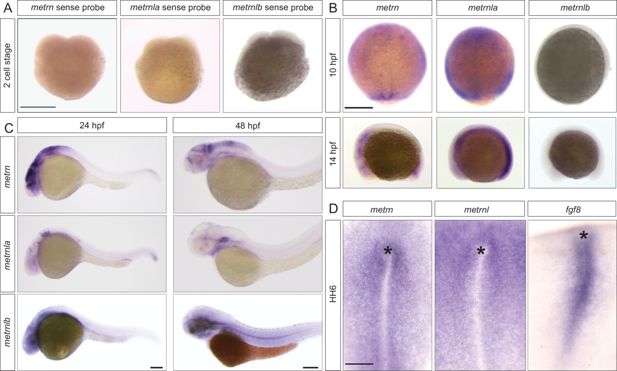
Fig. 3 - Supplemental 1 Metrns expression during early zebrafish and chick development. (A) No expression can be detected for metrn, metrnla, and metrnlb upon in situ hybridization using sense metrn, metrnla, and metrnlb riboprobes on two-cell stage wild type embryos. (B) Metrn expression at 10 hpf can be found in the leading edge of the shield and in the forming Kupffer’s vesicle as shown by in situ hybridization (dorsal view), and at 14 hpf, metrn transcripts can be detected in the area of the KV and the developing brain (later view). While metrnla at 10 hpf is expressed in the whole enveloping layer and developing midline as revealed here by in situ hybridization (dorsal view). At 14 hpf, metrnla transcripts can be found in the area of the forming KV and midline (later view). No expression can be detected for metrnlb at these stages. (C) From 24 hpf, all metrn genes are expressed in the developing central nervous system as shown by in situ hybridization (lateral views). (D) Dorsal view of metrn, metrnl, and fgf8 in situ hybridization in HH6 chick embryos reveals that metrn and metrnl are expressed around the Hensen’s node (*) and the primitive streak during chick early embryonic development. Scale bars: in (A, B, and D) 250 μm and in (C) 100 μm.