
Fig. 2
- ID
- ZDB-IMAGE-260408-24
- Genes
- Publication
- Chen et al., 2026 - Insulin-like growth factor signaling regulates zebrafish lymphatic-vessel development
- All Figures
- Figures for Chen et al., 2026
|
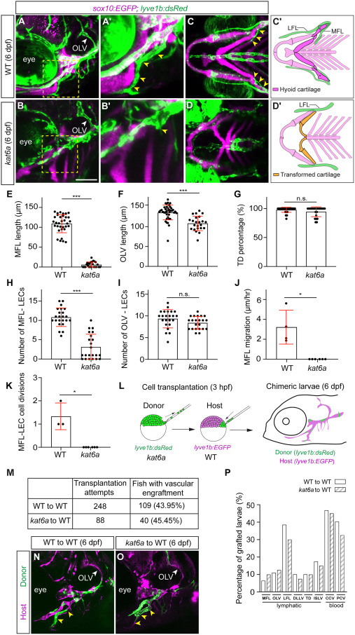
Fig. 2 kat6a is non-cell-autonomously required for MFL development (A and B) Confocal images of the lateral head, comparing the cartilage and lymphatic phenotypes between WT sibling (A) and kat6ab719 (B) lyve1b:dsRed (green) sox10:EGFP (magenta) larvae at 6 dpf. (A′ and B′) Magnified image regions are highlighted by the yellow dashed boxes in (A). Yellow arrowheads highlight the MFL. (C and D) The corresponding ventral view from (A) and (B). Note the underdeveloped MFL in kat6ab719 larvae (B′ and D). (C′ and D′) Diagrams illustrating hyoid cartilage and lymphatic (green) phenotypes from (C) and (D). (E–G) Quantification of the length of the MFL (E), the length of OLV (F), and the formation of TD (G) at 6 dpf, comparing kat6ab719 (n = 24) to WT siblings (n = 33). (H and I) Quantification of MFL-LECs (H) and OLV-LECs (I) at 6 dpf, comparing kat6ab719 (n = 22) to WT siblings (n = 23). (J and K) Quantification of MFL migration speed (J) and the number of MFL-LEC divisions from 4 to 5 dpf (K). Data were taken from 16- to 24-h time-lapse imaging experiments initiated at 4 dpf, comparing WT (n = 3–4) and kat6ab719 (n = 6). (L) Diagram showing the workflow of the cell-transplantation assay. (M) Table of vascular engraftment rates. (N and O) Confocal images of the lateral head at 6 dpf showing facial lymphatic engraftment of lyve1b:dsRed -expressing WT (N) or kat6ab719 (O) (green) donor cells into lyve1b:EGFP WT hosts (magenta). Yellow arrowheads highlight the MFL. (P) Graph displaying the proportion of grafted embryos with at least one donor cell engrafted in the indicated vessel. WT into WT (n = 109) with kat6ab719 into WT (n = 40). Data represent mean ± SD; ∗∗∗p < 0.001; ∗p < 0.05; ns, p > 0.05 from unpaired t test with Welch’s correction. CCV, common cardinal vein; DLLV, dorsal longitudinal lymphatic vessel; ISLV, intersegmental lymphatic vessel; LFL, lateral facial lymphatic vessel; MFL, medial facial lymphatic vessel; OLV, otolithic lymphatic vessel; PCV, posterior cardinal vein; TD, thoracic duct. Scale bar: 50 μm.