
Fig. 7
- ID
- ZDB-IMAGE-260408-29
- Genes
- Publication
- Chen et al., 2026 - Insulin-like growth factor signaling regulates zebrafish lymphatic-vessel development
- All Figures
- Figures for Chen et al., 2026
|
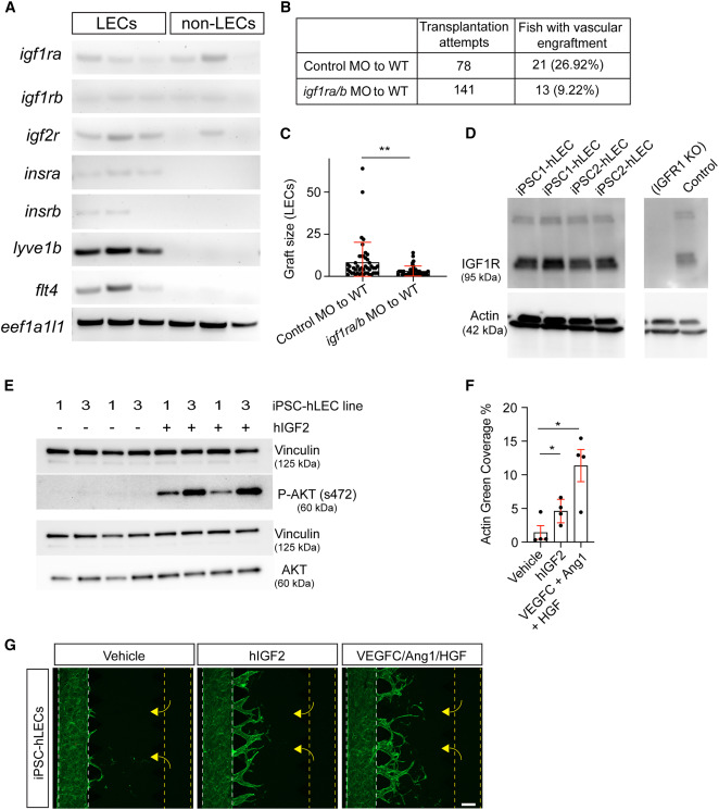
Fig. 7 IGF signaling is required cell autonomously for lymphatic-vessel growth (A) Gel electrophoresis of RT-PCR products demonstrating the expression of igfr family members in lymphatic endothelial cells (LECs) at 4 dpf. (B) Table of vascular engraftment rates. (C) Comparison of the average number of cells within each dsRed+ lymphatic graft between control MO-injected donors (n = 46) and igf1ra/igf1rb MO co-injected donors (n = 41). (D) Western blot confirming the expression of IGF1R in iPSC-hLECs (iPSC1 = ATCC-BYS0112, iPSC2 = ATCC-BXS0116). Lysates from mouse preadipocyte IGF1R KO cells and a control line are included as negative and positive controls, respectively.62 (E) Western blot demonstrating the activation of AKT (s472) in iPSC-hLECs following a 5-min incubation with 135 nM hIGF2 (iPSC1 = ATCC-BYS0112, iPSC3 = KOLF2.1J). (F) Quantitation of iPSC-hLEC migration in the microfluidic sprouting assay; n = 4 independent differentiations from two different iPSC lines (two differentiations each). (G) Confocal images of the in vitro microfluidic sprouting assay. Yellow dashed lines represent the channel containing either vehicle control or growth factors; white dashed lines represent the channel where iPSC-hLECs were seeded. Scale bar: 200 μm. Data represent mean ± SD; ∗∗p < 0.01; ∗p < 0.05; ns, p > 0.05 from one-way ANOVA with Dunnett’s multiple comparison tests for the significance assessment.