
|
Figure 4
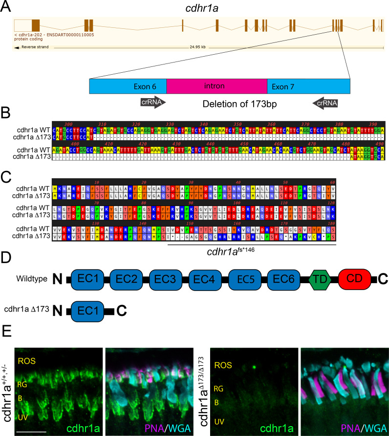
Construction and confirmation of the cdhr1a-/- line.
(A) Exon/intron diagram of cdhr1a. The intron 6-exon 6-intron 7 junction is highlighted and the approximate location of the crRNA (black arrows) is depicted. (B) Genomic nucleotide sequence from the cdhr1aΔ173 line highlighting the sequence location of the 173 bp deletion compared to WT sequence. (C) Amino acid sequence alignment between wildtype and the cdhr1aΔ173 line. Alignment indicates that the 173 bp deletion leads to a frameshift and an immediate premature stop codon at AA146. (D) Diagrammatic representation of the domain structure of WT cdhr1a protein vs the cdhr1aΔ173 allele. EC = cadherin domain, TD = transmembrane domain, CD = cytoplasmic domain. (E) Confocal microscopy of 5 dpf retinal cryosections from WT and cdhr1a-/- retinas stained with anti-cdhr1a antibody (green), PNA (red-green cones - magenta) and WGA (blue cones - teal). Cdhr1a signal is detected along photoreceptor outer segments in the wildtype and completely missing in cdhr1a-/-. ROS = rod outer segment, RIS = rod inner segment. Scale bar = 5 μm.