
Fig. 3
- ID
- ZDB-IMAGE-260723-41
- Publication
- Kaveh et al., 2026 - Flow-mediated endothelial remodeling and inflammation drive developmental vascular susceptibility in ldlr loss of function
- All Figures
- Figures for Kaveh et al., 2026
|
Fig. 3
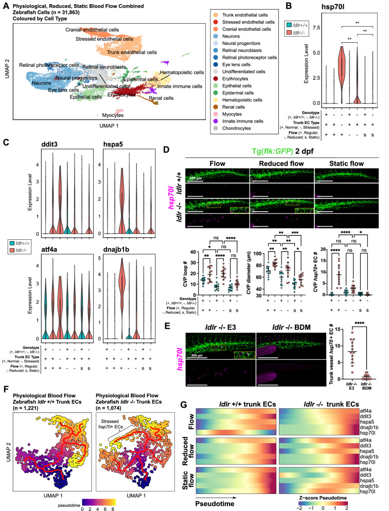
Endothelial hsp70 upregulation and altered remodeling angiogenesis are mediated by blood flow in ldlr-/- embryos.
A Combined cluster UMAP of 31,863 single cells showing annotated cell populations from embryonic ldlr + / + and ldlr-/- zebrafish under physiological, reduced (0.4 ng tnnt2a morpholino) and static (2 ng tnnt2a morpholino) blood flow conditions. Cells were obtained from pooled embryos (n = 200 zebrafish per group) processed in a single batch. B Violin plot of normalized hsp70l expression in trunk and stressed endothelial cells across genotypes and flow conditions. Each violin represents single-cell expression values, the central line indicates the median, and the shape represents the kernel distribution. Two-tailed Wilcoxon rank sum test and Bonferroni’s multiple comparison test used. ** p < 0.01. C Violin plots of normalized cell stress response expression (ddit3, hspa5, atf4a and dnajb1b) in trunk and stressed endothelial cells across genotypes and flow conditions. D Confocal microscopy of hsp70l fluorescence in situ RNA hybridization (FISH) stained trunk vasculature in ldlr + /+ and ldlr-/- zebrafish under physiological, reduced and static blood flow at 2 dpf. The inset shows CVP hsp70 + endothelial cells adjacent to CVP loops (top). Caudal venous plexus (CVP) metrics: loop number, diameter (μm) and hsp70+ endothelial cell number in ldlr + /+ (n = 10–12) and ldlr-/- zebrafish (n = 10–12) across flow conditions. Flow ldlr + / + vs flow ldlr-/- (loop number: p = 0.0086, diameter: p = 0.0030, hsp70 + number: p < 0.0001). Reduced flow ldlr + /+ vs reduced flow ldlr-/- (loop number: p < 0.0001, diameter: p = 0.0039, hsp70 + number: p = 0.2918). Static flow ldlr + /+ vs static flow ldlr-/- (loop number: p = 0.1083, diameter: p = 0.0226, hsp70 + number: p = 0.7410). Flow ldlr + / + vs reduced flow ldlr + / + (loop number: p = 0.0105, diameter: p = 0.0048, hsp70+ number: p = 0.4621). Reduced flow ldlr + /+ vs static flow ldlr + /+ (loop number: p = 0.7196, diameter: p = 0.0046, hsp70+ number: p = 0.6153). Flow ldlr-/- vs reduced flow ldlr-/- (loop number: p = 0.7196, diameter: p = 0.0002, hsp70+ number: p < 0.0001). Reduced flow ldlr-/- vs static flow ldlr-/- (loop number: p < 0.0001, diameter: p = 0.0046, hsp70 + number: p = 0.0218). 3 independent experiments. Data are mean ± s.d. Two-way ANOVA and Holm-Sidak’s multiple comparison used (bottom). E Confocal microscopy of hsp70l FISH-stained trunk vasculature in ldlr-/- zebrafish exposed to E3 (control) medium or 10 mM BDM (1-2 dpf). Number of trunk vessel hsp70 + endothelial cells in ldlr-/- zebrafish exposed to E3 control medium (n = 13) or 10 mM BDM (n = 13); p < 0.0001; 3 independent experiments. Data are mean ± s.d. Paired two-tailed t test was used (right). F Pseudotime trajectory UMAP of trunk endothelial cells in ldlr + /+ (left) and ldlr-/- (right) zebrafish under physiological blood flow. Trajectory and lineage path of stressed hsp70 + endothelial cells is indicated in ldlr-/- zebrafish (absent in ldlr + /+ zebrafish) with flow. G Pseudotime z-score heatmaps of cell stress response genes (atf4a, ddit3, hspa5, dnajb1b and hsp70l) in ldlr + / + (left) and ldlr-/- (right) trunk endothelial cells under physiological (top), reduced (middle) and static (bottom) blood flow. Source data are provided as a Source Data file.