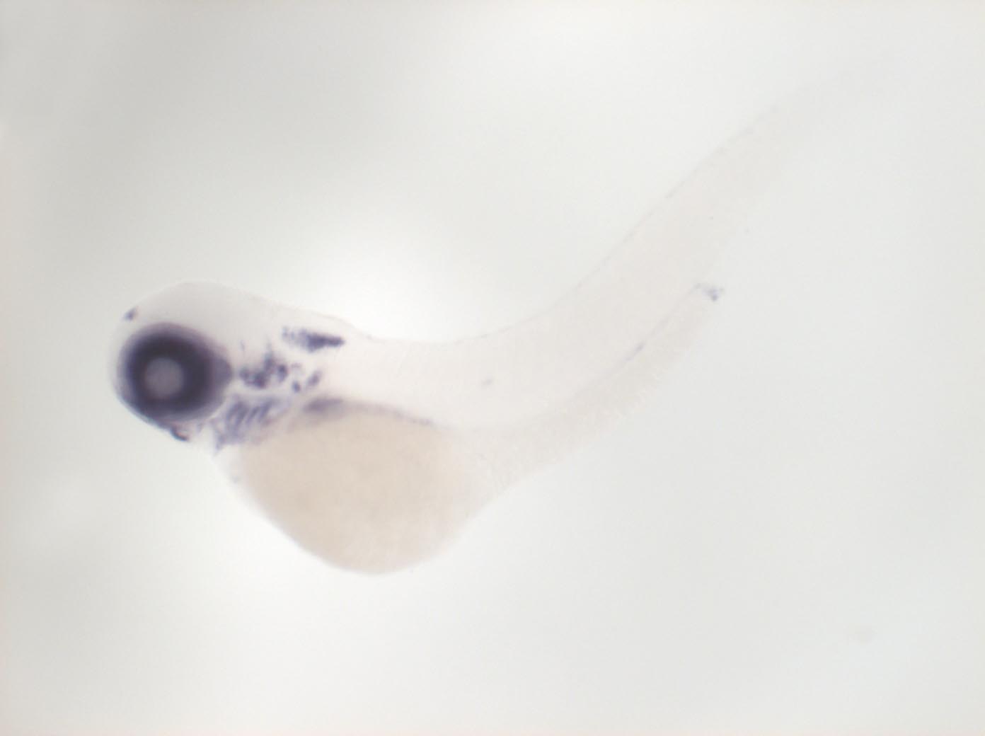
Image


                        Figure Caption


Fig. 6 Expressed in retina (ganglion and intermediate cell layer), epiphysis, otic vesicle, pharyngeal arches 3-7, dorsal anterior spinal cord, hypaxial muscles, pectoral fin
                
                    
                        Orientation


                | Preparation | Image Form | View | Direction | 
| whole-mount | still | side view | anterior to left | 
                
                    
                        Figure Data


        