
Fig. 3
- ID
- ZDB-FIG-090504-55
- Publication
- Heisenberg et al., 1997 - The function of silberblick in the positioning of the eye anlage in the zebrafish embryo
- Other Figures
- All Figure Page
- Back to All Figure Page
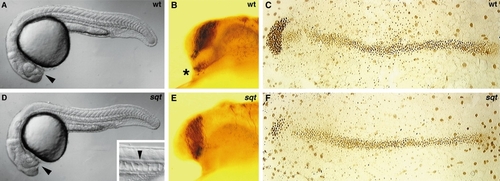
In sqt embryos, the eyes are fused anteriorly and the ventral diencephalon as well as the underlying prechordal plate is reduced. (A, D) Side view of a wild-type (A) and sqt (D) embryo at 24 hpf. (B, E) The axonal scaffold stained with an anti-acetylated tubulin antibody in wild-type (B) and sqt (E) embryos at 24 hpf. (C, F) The axial mesendoderm stained with an anti-Fkd2 antibody in wild-type (C) and sqt (F) embryos at bud stage. Arrowheads in A and D point to the position of the optic stalks. Small picture in the bottom right corner of D shows the presence of a floorplate (arrowhead) in sqt mutants at 24 hpf. Asterisk in B demarcates the position of the nucleus of the tract of the postoptic commissur. Side (A, B, D, E) and dorsal (C, F) views, anterior to the left. |
| Fish: | |
|---|---|
| Observed In: | |
| Stage Range: | Bud to Prim-5 |
Reprinted from Developmental Biology, 184(1), Heisenberg, C.P. and Nüsslein-Volhard, C., The function of silberblick in the positioning of the eye anlage in the zebrafish embryo, 85-94, Copyright (1997) with permission from Elsevier. Full text @ Dev. Biol.