
Fig. 2
- ID
- ZDB-FIG-110407-39
- Publication
- Carvalho et al., 2011 - A high-throughput screen for tuberculosis progression
- Other Figures
- All Figure Page
- Back to All Figure Page
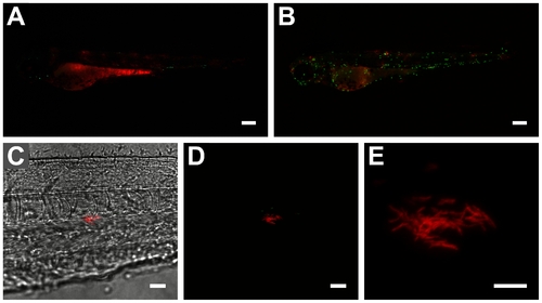
Effect of yolk sac co-injection of Pu.1 morpholino and M. marinum on bacterial localization and proliferation within embryos. (A and B) 3 day-old infected mpx-gfp transgenic embryos (A) with and (B) without Pu.1 morpholino (scale bar: 250 μm). Greater numbers of (extracellular) bacteria throughout body of morphant embryo seen in A contrast with lower amount of more localized (phagocytosed) bacteria seen in B. Very low number of mpx-gfp labelled neutrophils in A confirms Pu.1 morpholino effect. (C) Bright-field/fluorescence overlay and (D) confocal z-stack of mag49-GFP/mCherry bacteria in body of 2 dpi embryo (scale bar: 25 μm). Red-fluorescent bacteria form a cording structure adjacent to a few cells containing green-fluorescent (mag49-activated) bacteria. Lack of green fluorescence in cording bacteria indicates no phagocytosis by macrophages and extracellularity. (E) Close-up (digital zoom: 5.2) of cording structure formed by extracellular bacteria (scale bar: 10 μm; only red channel shown). |
| Fish: | |
|---|---|
| Condition: | |
| Knockdown Reagent: | |
| Observed In: | |
| Stage: | Long-pec |