
Fig. 2
- ID
- ZDB-FIG-111223-22
- Publication
- Herwig et al., 2011 - Distinct Cellular Mechanisms of Blood Vessel Fusion in the Zebrafish Embryo
- Other Figures
- All Figure Page
- Back to All Figure Page
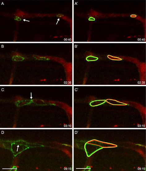
Anastomosis by Cord Hollowing Still pictures from Movie S2 showing a time-lapse of a fli:GAL4FF; UAS:RFP; UAS:EGFP-ZO1 embryo (total length: 9:20). (A–C) A tip cell has established contact with an adjacent tip cell (right arrow) and has contact with a stalk cell (left arrow), which results in two loops of EGFP-ZO1 (A). These loops extend and eventually meet (B and C). (A′–D′) shows the junctional outline of participating cells (yellow: “central” cell, green: stalk cell, red: DLAV cell moving in from posterior [right]). The two initial cell contacts are shown in yellow/green and yellow/red, respectively. When the stalk cell and the adjacent DLAV cell meet, they establish a new contact with “green and red” junctions (arrow in D, green and red line in D′). Scale bar in all pictures represents 20 μm. |