
Fig. 5
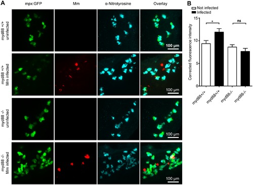
Increased tyrosine nitration following Mm infection is dependent on MyD88. (A) Fluorescent micrographs of confocal images showing the overlap between mpx:GFP and anti-nitrotyrosine staining in myd88+/+ or myd88-/- embryos. (B) Example fluorescence confocal z-stacks of the caudal vein region of myd88+/+ or myd88-/- embryos stained with anti-nitrotyrosine antibody, imaged at 1 dpi in the presence or absence of Mm infection. (C) Corrected fluorescence intensity levels of anti-nitrotyrosine antibody confocal z-stacks of equal size 1 day after injection with Mm in myd88+/+ or myd88-/- embryos. Data shown are mean ± SEM, n = 30 cells from 5 embryos representative of 2 independent experiments. |
| Gene: | |
|---|---|
| Antibody: | |
| Fish: | |
| Condition: | |
| Anatomical Term: | |
| Stage: | Long-pec |