|
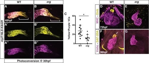
SHF-derived VC and smooth muscle development is impaired in the <italic>crg</italic> mutants.(A-B”) Representative images of hearts from photoconverted WT sibling and crg mutant myl7:NLS-KikGR embryos at 48 hpf. The arterial poles (brackets) are to the right. (C) Quantification of later-differentiating VCs (Yellow+/Purple- cells) (n = 10 for WT and crg mutants). (D,E) Confocal images of IHC for MHC and Elnb in WT and crg mutant embryos at 72 hpf. n = 10 WT and n = 10 crg mutants embryos examined. (F,G) Confocal images of DAF-2DA staining coupled with IHC for Vmhc in WT sibling and crg mutant embryos at 96 hpf. n = 10 WT and n = 10 crg mutants embryos examined. Images in D-G are frontal views with anterior up. Arrows indicate Elnb and DAF-2DA staining of the bulbous arteriosus.
|