|
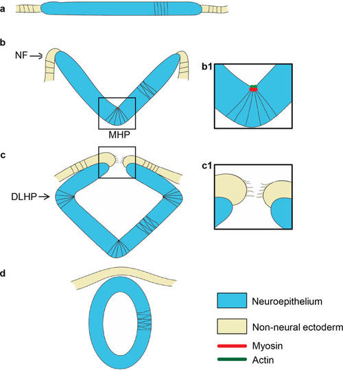
Neurulation in amniotes. Cross-sectional illustration of stages of neurulation in amniotes.a The neural plate and adjacent non-neural ectoderm. b Medial hingepoint formation shapes the neural groove and elevates the neural folds. b1 Illustration of medial hingepoint cells that are apically constricted and enriched for actomyosin at their apex. c Dorso-lateral hingepoint formation brings the neural folds in close apposition. c1 Filopodial extensions establish contact between neural fold cells across the midline. In the mouse forebrain, the first contact is established between neuroectodermal cells. d The neural folds fuse medially, separating the epidermis from the neural tube. DLHP dorso-lateral hingepoint, MHP medial hingepoint, NF neural fold.
|