
Fig. 4
- ID
- ZDB-FIG-220810-20
- Publication
- Chen et al., 2022 - Leukocyte invasion of the brain after peripheral trauma in zebrafish (Danio rerio)
- Other Figures
- All Figure Page
- Back to All Figure Page
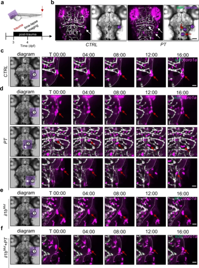
a Experimental setup. b Active leukocytes (white arrow) were distributed in the brains of zebrafish with or without peripheral trauma. Scale bar, 40 µm. c Time-lapse light-sheet imaging showed the active leukocytes (red arrow) around the zebrafish brain in CTRL. Scale bar, 20 µm. d Time-lapse light-sheet imaging showed active leukocytes (red arrow) around the brain (1), inside the brain (2), and invading the brain (3) at 4 dpf. Scale bar, 20 µm. e Time-lapse light-sheet imaging showed the active leukocytes (red arrow) around the brain in il1bMut zebrafish. Scale bar, 20 µm. f Time-lapse light-sheet imaging showed the active leukocytes (red arrow) around the brain in il1bMut zebrafish at 4 dpf. Scale bar, 20 µm. |
| Fish: | |
|---|---|
| Condition: | |
| Observed In: | |
| Stage: | Day 4 |