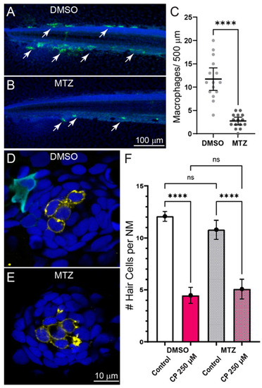
Depleting macrophages does not affect cisplatin induced neuromast hair cell loss. (A–C) Selective depletion of macrophages in double transgenic fish (gl25Tg, Tg(mpeg1.1:GAL4FF); c264Tg, Tg(UAS-E1B:NTR-mCherry). (A,B) Representative images of macrophage distribution (indicated with arrows) within the posterior-most 500 µm of the spinal column of fish treated with for 24 h with 0.1% DMSO (A) or 10 mM MTZ (B). (C) MTZ-treated fish showed a significant depletion of macrophages relative to DMSO-treated control (Unpaired t-test; **** p < 0.0001). Bars = Mean w/95% CI. Data obtained from 16 fish/treatment group. (D,E) Single z-section images taken from confocal stacks of neuromasts in fish following treatment with 250 µM cisplatin. Macrophages are labeled with YFP (cyan), hair cells with an antibody to Otoferlin (HCS-1; yellow) and all cell nuclei are labeled with DAPI (blue). (F) Quantification of hair cell loss following exposure to 250 µM cisplatin and 24 h recovery. Significant hair cell loss following cisplatin was observed in both DMSO and MTZ treatment groups relative to controls (Tukey’s multiple comparisons test, **** adjusted p < 0.0001), but no difference in cisplatin-induced hair cell loss was observed between DMSO and MTX treatment groups (adjusted p = 0.6415) Bars = Mean w/95% CI. n = 37–39 fish per treatment group, N = 3 trials.
|
