
Figure 5 - figure supplement 2
- ID
- ZDB-FIG-230123-24
- Publication
- Sabharwal et al., 2022 - Genetic therapy in a mitochondrial disease model suggests a critical role for liver dysfunction in mortality
- Other Figures
-
- Figure 1
- Figure 2
- Figure 2 - figure supplement 1
- Figure 2 - figure supplement 2
- Figure 2 - figure supplement 3
- Figure 3
- Figure 3 - figure supplement 1
- Figure 4
- Figure 4 - figure supplement 1
- Figure 5
- Figure 5 - figure supplement 1
- Figure 5 - figure supplement 2
- Figure 6
- Figure 7
- Figure 7 - figure supplement 1
- Figure 8
- All Figure Page
- Back to All Figure Page
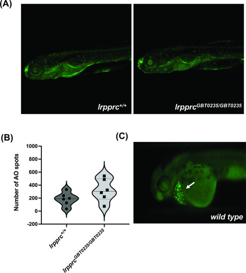
lrpprc homozygous mutants do not display neuronal necrosis. (A) Representative images of 6 dpf wild-type and lrpprcGBT0235/GBT0235 mutants. Background neuronal necrosis was observed in the wild type as well as lrpprc homozygous siblings (magnification- 5×). (B) Individual spots were quantified in the neuronal region of interest across a series of images (blinded images) obtained from both genotypes and the number of such particle counts was not significant (p-value = 0.1797). Each individual data point represents a single embryo. p-Values were determined using the Mann-Whitney U test. (C) Hatching gland displaying programmed apoptosis during organogenesis at 2 dpf zebrafish embryo (Positive control for the AO assay; Figure 5—source data 1). |