|
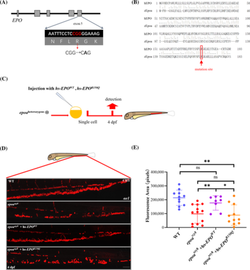
Human EPO variants assessment in epoaszy8. (A) Location of the R150Q EPO mutation in genomic sequence. The mutation is highlighted and marked with a red arrow in the sequence below. (B) Amino acid sequence alignment of human EPO and zebrafish Epoa, location of the R150Q EPO mutation is marked with a red line in the sequence. (C) Schematic diagram of the validation of epoaszy8 model using human DBAL related EPO mutation. The hsEPOWT, hsEPO530G>A mRNA were injected into one cell of the embryos from epoaheterozygous intercross. (D) Antibody staining of αe1+ cells in CHT of 4-dpf larvae injected with human hsEPOWT mRNA, hsEPO530G>A mRNA. (E) Statistical significance was determined using a two-sample Student's t-test, n ≥ 8, mean ± SD, ns: not significant, *p < .05 and **p < .01. Scale bars: 50 μm.
|