|
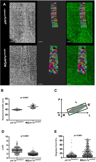
MZptk7audm400 mutants display defective convergent extension of notochord precursor cells. (A) Multiphoton image (left) of ptk7audm400/+ (top) and MZptk7audm400 embryos (bottom) at 14 hpf. Dorsal views of 2D renderings of notochord cells are shown in the middle panel and the overlap on the right. Vacuolated cells are colored artificially. Scale bars: 30 µm. (B) The notochord is significantly wider in MZptk7audm400 (n=12) compared with their ptk7audm400/+ siblings (n=9). P-value was calculated using two-tailed Welch's t-test. (C) A schematic of the method used to measure length-to-width ratio (LWR) and cell angle relative the mediolateral (ML) axis. A, anterior; P, posterior. (D,E) LWR is significantly lower (D) and the M-L orientation is significantly defective (E) in MZptk7audm400 (3 embryos, n=331) compared with their ptk7audm400/+ siblings (3 embryos, n=303). P-value was calculated using two-tailed Mann–Whitney U-test.
|