FIGURE

Fig. 3

ID
ZDB-FIG-241213-38
Publication
Xu et al., 2024 - PDGFRA is a conserved HAND2 effector during early cardiac development
Other Figures
All Figure Page
Back to All Figure Page
Fig. 3

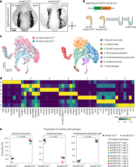
scRNA-seq analysis of hand2 reporter-expressing cells in WT and hand2 mutant embryos.

a, Maximum intensity projections of confocal images of 20 hpf TgBAC(hand2:EGFP) expression in hand2 FLD+/+ and hand2 FLD−/− sibling embryos. The proportion of embryos matching the image shown is indicated in the top right corner of each image. The scale bar applies to all images. b, Schematic of the experimental protocol; the transcriptomes of 3,900 and 3,836 individual hand2 reporter-expressing cells from 24 hpf hand2 FLD−/− and hand2 FLD+/? sibling embryos, respectively, were sequenced. c, Left, UMAP representation of the cells from hand2 FLD−/− (blue) and hand2 FLD+/? (pink) sibling embryos. Right, UMAP of the data clustered by the Leiden algorithm. d, Heat map of the top 7 DEGs in each cell cluster. A list of the DEGs in each cell cluster can be found in Supplementary Table 2. e, Proportional contribution of cardiac precursors, cardiomyocytes and endothelial/endocardial cells in 24 hpf hand2 FLD+/? and hand2 FLD−/− sibling embryos generated by Scanpro’s bootstrapping method. Panel b created with BioRender.com.

Expression Data

Expression Detail
Antibody Labeling
Phenotype Data

Phenotype Detail
Acknowledgments
This image is the copyrighted work of the attributed author or publisher, and ZFIN has permission only to display this image to its users. Additional permissions should be obtained from the applicable author or publisher of the image. Full text @ Nat Cardiovasc Res