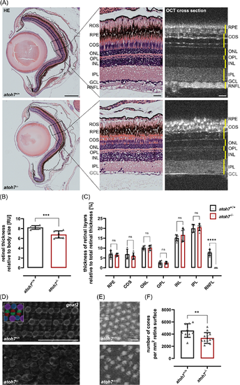
atoh7 Mutant retinae show reduced thickness and structural defects. (A) Hematoxylin and eosin (H&E) stainings of wildtype and mutant animals. Overall, retinal morphology is similar in wildtype as well as mutants and individual nuclear and plexiform layers can be distinguished (left panel). However, detailed analysis (middle panel) revealed that mutant retinae miss the retinal nerve fiber layer (RNFL). Additionally, the cone outer segments (COS) appear to be less organized and the rod outer segments (ROS) are shorter in mutant specimens. Optical coherence tomography (OCT) cross sections in live animals confirmed these observations (right panel). (B) Quantification showed that the relative retinal thickness of mutant fish (compared to body size) is significantly reduced. (C) Quantifications of the relative thicknesses of individual layers (as percentage of whole retinal thickness, measured layers depicted in yellow in A) showed no significant differences except for the RNFL. (D) Gnat2 staining of cone outer segments in retinal flat mounts reveals defects in the regular cone mosaic (colored scheme on the upper left) in mutant retina. (E) Cone photoreceptor mosaic in wildtype and mutant retinae in en face projections calculated from OCT cross sectional scans. (F) Quantification of the number of cones in the OCT en face projections revealed a significant reduction in mutant retinae. Scale bars represent 250 µm in overviews, 25 µm in magnifications (A) and 50 µm in flat mounts (D, E). Statistics: All data are represented as mean ± SD, unpaired t-test (B, F), t-test with multiple comparisons (C), *P < 0.05; **P < 0.01; ***P < 0.001, or ****P < 0.0001. COS, cone outer segments; GCL, ganglion cell layer; INL, inner nuclear layer; IPL, inner plexiform layer; ONL, outer nuclear layer; OPL, outer plexiform layer; RNFL, retinal nerve fiber layer; ROS, rod outer segments; RPE, retinal pigment epithelium.
|
