|
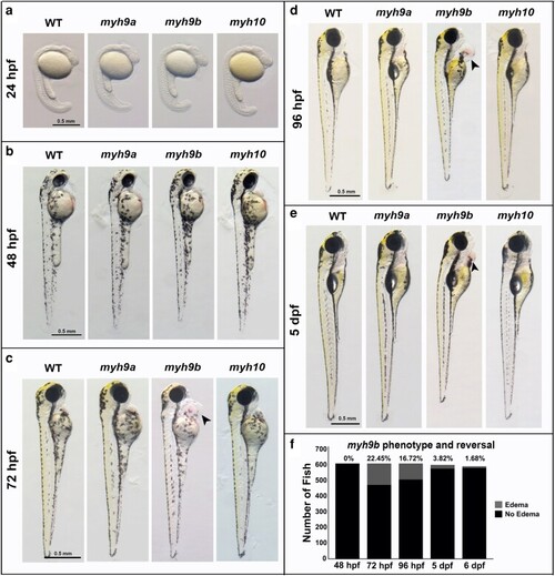
myh−/− mutant phenotypes between 24 hpf and 5 dpf. a–e) Representative bright-field images of myh9a−/−, myh9b−/−, and myh10−/− homozygous mutants compared to WT at 24 hpf, 48 hpf, 72 hpf, 96 hpf, and 5 dpf. Arrowheads indicate location of pericardial edema in myh9b−/− mutants. f) Quantification of myh9b pericardial edema phenotype development and reversal from 48 hpf to 6 dpf in offspring generated from an myh9b+/− parent incross. The χ2 analyses determined that at 72 hpf the edema phenotype development is Mendelian, and at 96 hpf and beyond it is no longer Mendelian. Scale bars for all images = 0.5 mm. n = 610 for phenotype development and reversal quantification in f.
|