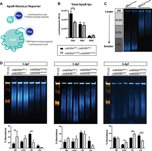
creb3l3ac802/c802;creb3l3bc803/c803 mutants have fewer and larger ApoB-lps at 3 dpf. A: Schematic of LipoGlo reporter on differently sized ApoB-lps. B: Quantification of total ApoB-lps in creb3l3ac802;creb3l3bc803 mutants compared to double heterozygous siblings (creb3l3ac802/+;creb3l3bc803/+) from 3 to 6 dpf. All fish used in these experiments were heterozygous for the ApoBb.1-NanoLuc reporter. Three independent experiments, 8–12 individuals per genotype. One-way ANOVA, ∗∗∗∗P < 0.001. C: ApoB-lp particle size distributions of 6 dpf ldlrasd52 mutant and apoc2sd38 mutant homogenate for comparison. D: Native PAGE shows size distribution of ApoB-lp in creb3l3 double homozygous mutants from 3 to 6 dpf compared to creb3l3 double heterozygous siblings. Gel images are a composite of chemiluminescent signal (LipoGlo, blue) and fluorescent (DiI-LDL, orange) exposures. DiI-labeled human LDL sample (L3482, Thermo Fisher Scientific) is run in the first lane of each gel as a migration standard. Human Dil-LDL migrates more slowly than NanoLuc-labeled LDL due to the addition of the DiI label. Quantification of luciferase signal on Native PAGE at 3, 5, and 6 dpf by percent abundance of signal within a lane. Gels images are representative of three independent experiments. One-way ANOVA with Tukey’s multiple comparisons. N = 3 clutches; 3–12 individuals per genotype per clutch. ∗∗P < 0.01; ∗∗∗P < 0.005; and ∗∗∗∗P < 0.001. ApoB-lp, apolipoprotein-B–containing lipoprotein; Creb3l3, cAMP-responsive element-binding protein 3–like 3; DiI-LDL, DiI-labeled LDL; dpf, days post fertilization; IDL, intermediate-density lipoprotein; ZM, zero mobility.
|
