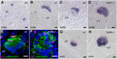
Mutual repression by frizzled9b and wnt4 signaling patterns tubule junctions. (A) fzd9b is expressed in single nephron progenitor cells prior to aggregation. (B,C) After acute injury, fzd9b expression persists in new cell aggregates (B) but is downregulated in cells adjacent to the distal tubule (C; DT, dotted outline), a domain of high canonical Wnt signaling (asterisk in C). (D) fzd9b expression in wnt4 mutant new nephrons escapes downregulation and remains uniformly expressed in new nephrons. (E) Expression of the canonical Wnt reporter Tg(tcflef:dGFP) is restricted to new nephron cells adjacent to the distal tubule (DT) and is not expressed in cells several cell diameters away (asterisk). (F) Mutation in fzd9b results in expanded expression of Tg(tcflef:dGFP) where the entire new nephron can show GFP expression (asterisk). (G) wnt4, an endogenous canonical Wnt target gene, is expressed in new nephron cells adjacent to the distal tubule. (H) Mutation in fzd9b significantly expands wnt4 expression. Blue fluorescence in E and F indicates Hoechst-stained nuclei. Scale bars: 20 µm.
|
