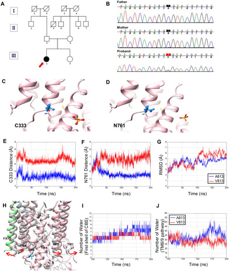
Family pedigree and genetic analysis and structural changes of the ATP1A3-p.A813V mutation. (a) Genetic lineage of a rapid-onset dystonia-parkinsonism (RDP) patient family. The inheritance pattern, showing the heterozygous mutation carrier alongside unaffected parents. (b) DNA sequence electropherograms (father, mother, proband) revealing wild-type sequence of the parents and the de novo mutation c.2438C>T, A813V (p.Ala813Val) in the proband. (c-d) wild-type (WT) ATP1A3 structure illustrating stable interactions of A813 with C333 (TM4) and N761 (TM5), which preserves TM helix packing. (e-f) ATP1A3-p.A813V mutant structure (red) exhibits increased Cα distances between V813 and interacting residues (A813V-C333: 4.34 Å → 6.59 Å; A813V-N761: 4.76 Å → 6.66 Å) compared with the wild type (blue), suggesting destabilization due to the bulkier valine residue. (g) Root-mean-square distance (RMSD) analysis shows that while the WT remains stable, the mutant deviates after 100 ns, indicating progressive structural instability. (h) Superimposition of the initial structure (light gray color) and the representative structure of the most populated cluster of A813V mutant MD trajectory. ATP1A3, ATP1B1, FXYD, and V813 are colored pink, light blue, light green, and blue, respectively. Number of water molecules in the first shell of CBS (i) and TM6/9 pathway (j). Blue and red lines are WT and mutant, respectively
|
