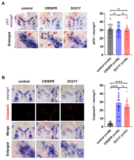
Loss of Pla2g6 induces apoptosis of neuronal precursors without affecting proliferation. Embryos at 10 hpf were flat-mounted after in situ hybridization to facilitate visualization and quantification of signal-positive cells. (A) Proliferation analysis. neurog1 in situ hybridization was combined with immunodetection of phosphorylated histone H3 (pH3). Representative images are shown on the left, with quantification of pH3-positive cells within the neurog1-positive region shown on the right. The number of proliferating neuronal precursors was not significantly different between control and Pla2g6-deficient embryos. (B) Apoptosis analysis. Co-labeling of neurog1 transcripts with cleaved caspase-3 identified apoptotic neuronal precursors. Representative images are shown on the left, with quantification of caspase-3-positive cells within the neurog1-positive region shown on the right. The bottom rows in panels A and B show enlarged views of the boxed regions (red dashed rectangles), with white arrowheads indicating caspase-3-positive signals. Pla2g6-deficient embryos exhibited a significant increase in apoptotic neuronal precursors compared with controls. Results composed of three independent biological replicates, with 7–10 embryos analyzed per experiment. Individual data points represent cell counts obtained from single embryos. Data are presented as mean ± SEM. Statistical significance was determined using one-way ANOVA with Tukey’s pairwise comparison; ****, p < 0.0001 ns, not significant.
|
