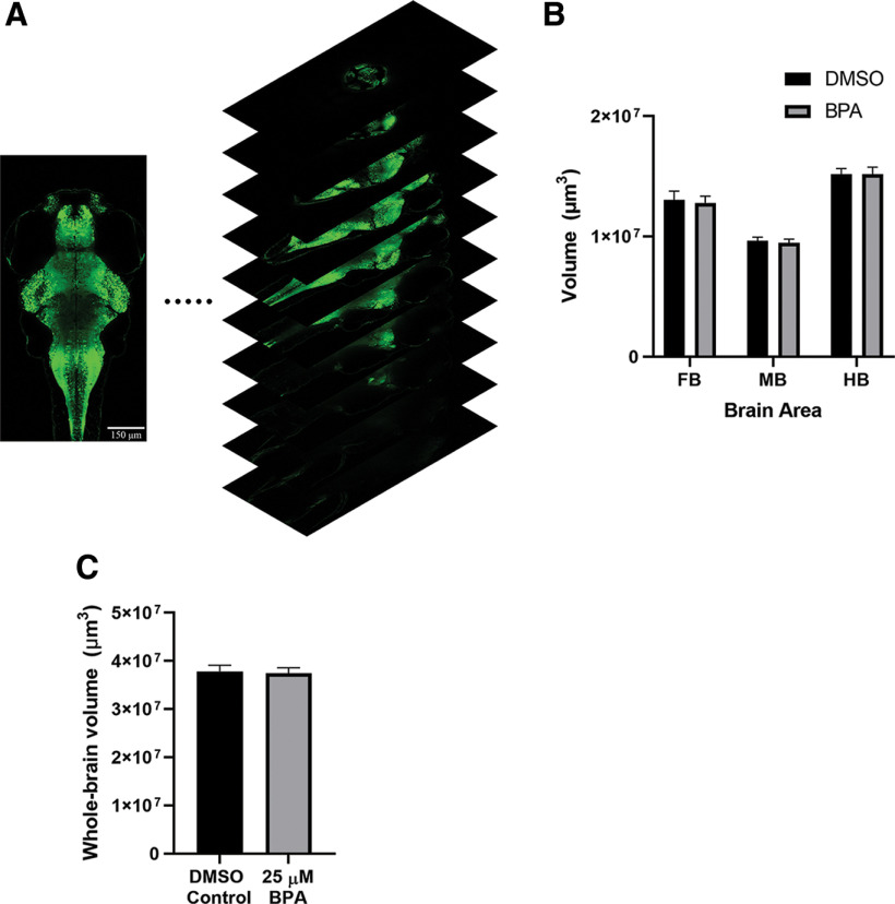
Brain volume was not changed in zebrafish larvae by BPA exposure. A, 3D reconstruction of a larval brain. Sample confocal images of optical sections of a brain exposed to DMSO. Scale bar: 150 μm. B, Mean volumes of the forebrain (FB), midbrain (MB), and hindbrain (HB) in BPA-exposed (BPA group, n =15) fish compared with the vehicle-exposed fish (DMSO group, n =15). A two-way ANOVA revealed no significant interaction or group effect (interaction, F(2,84) = 0.03; p =0.75: group, F(1,84) = 0.10; p =0.97. C, Mean volume of the whole brain in BPA-exposed fish (BPA group, n =15) and vehicle-exposed fish (DMSO group, n =15). The difference between the two groups was not significant (t(28) = 0.23, p =0.82).
This image is the copyrighted work of the attributed author or publisher, and
ZFIN has permission only to display this image to its users.
Additional permissions should be obtained from the applicable author or publisher of the image.
Full text @ eNeuro
Your Input Welcome
Thank you for submitting comments. Your input has been emailed to ZFIN curators who may contact you if
additional information is required.
Oops. Something went wrong. Please try again later.