IMAGE

Figure 2.

ID
ZDB-IMAGE-231031-17
Source
Figures for Liu et al., 2023
Image
Figure Caption

Figure 2.

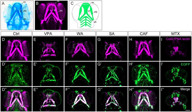
Craniofacial anomalies were identified in teratogen-treated sox10:EGFP zebrafish embryos at 96 hpf. (A, B) Craniofacial cartilage stained with Alcian blue (A) and anti-coll2 antibody and PNA lectin (B). (C) Schematic illustration of craniofacial cartilage elements. m, Meckel’s cartilage; p, palate; pq, palatoquadrate; ch, ceratohyal. (D–I″) sox10:EGFP embryos were treated with the following teratogens and displayed craniofacial anomalies. (D–D″) Ctrl, E3 control. (E–E″) VPA, valproic acid (15 mM). (F–F″) WAF, warfarin (30 mM). (G–G″) SA, salicylic acid (300 mM). (H–H″) CAF, caffeine (500 mM). (I–I″) MTX, methotrexate (200 mM). Immunohistochemical staining of craniofacial cartilage was performed with anti-coll2 antibody and PNA lectin (D–I), and anti-GFP antibody (D′–I′) following the teratogen exposure. Anti-GFP staining was co-merged with the cartilage staining (D″–I″). Control: n = 25, VPA: n = 18, WAF: n = 23, SA: n = 16, CAF: n = 31, MTX: n = 26. Bracket indicates the lower jaw of zebrafish. Dashed lines indicate outline of the palate. All images were taken from ventral view. m, Meckel’s cartilage; p, palate; pq, palatoquadrate. Scale bar: 100 µm.

Acknowledgments
This image is the copyrighted work of the attributed author or publisher, and ZFIN has permission only to display this image to its users. Additional permissions should be obtained from the applicable author or publisher of the image. Full text @ Toxicol. Sci.